amino acids

Essential amino acids in adult humans: lysine, leucine, isoleucine, methionine, phenylalanine, threonine, tryptophan, and valine.
Additional amino acids essential to children: histidine and arginine.

amino acid chartalaninearginineasparagineaspartic acidcysteineglutamic acidglutamineglycinehistidineisoleucineleucinelysineN-formylmethioninemethioninephenylalanineprolineserinethreoninetryptophantyrosinevaline

External links: Alanine Arginine Asparagine Aspartic acid Cysteine Glutamic acid Glutamine Glycine Histidine Isoleucine Leucine Lysine Methionine Phenylalanine Proline Serine Threonine Tryptophan Tyrosine Valine Essential amino acid Protein Peptide Genetic code Isoleucine and valine biosynthesis Leucine biosynthesis

amino acid chart

alanine


Non-polar amino acid Ala A RNA code: GCU, GCC, GCA, GCG

arginine


Basic amino acid Arg R RNA code: CGU, CGC, CGA, CGG, AGA, AGG

asparagine


Polar amino acid Asn N RNA code: AAU, AAC

aspartic acid


Acidic amino acid Asp D RNA code: GAU, GAC

cysteine


Polar amino acid Cys C RNA code: UGU, UGC

glutamic acid


Acidic amino acid Glu E RNA code: GAA, GAG

glycine


Non-polar amino acid Gly G RNA code: GGU, GGC, GGA, GGG

glutamine


Polar amino acid Gln Q RNA code: CAA, CAG

histidine


Basic amino acid His H RNA code: CAU, CAC

Histidine's imidazole side chain has a hydrogen-bound nitrogen atom that is slighty acidic and another nitrogen that is slightly basic. The almost neutral pKa of histidine ensures that its charge will change in response to relatively small shifts in cellular pH.

Histidine functions as a co-ordinating ligand in metalloproteins, and at the catalytic site of some enzymes. In catalytic triads, the basic nitrogen of histidine extracts a proton from serine, threonine or cysteine, activating the nitrogen as a nucleophile. In a histidine proton shuttle, histidine rapidly shuttles protons by abstracting a proton onto the basic nitrogen to generate a positively-charged intermediate before passing a proton from the acidic nitrogen. Carbonic anhydrases employ the histidine proton shuttle to rapidly shuttle protons away from a zinc-bound water molecule to quickly regenerate the active form of the enzyme.

isoleucine

Non-polar amino acid Ile I RNA code: AUU, AUC, AUA

leucine

Non-polar amino acid Leu L RNA code: UUA, UUG, CUU, CUC, CUA, CUG

lysine


Basic amino acid Lys K RNA code: AAA, AAG

N-formylmethionine


N-Formylmethionine (fMet) is an amino acid that is found in all living cells. It is a derivative of the amino acid methionine, in which a formyl group has been added to methionine's amino group (catalyzed by the enzyme transformylase when Met is attached to tRNA.fMet, but not to tRNA.Met).

N-Formylmethionine plays a crucial role in the protein biosynthesis of bacteria, archaea, mitochondria and chloroplasts. Bacteria utilize N-formylmethionine (f-Met) rather than methionine at the N-terminal of the elongating polypeptide chain.

Compare to methionine.

f-Met amino acid RNA code: AUG tRNA.fMet & tRNA.Met

methionine


Non-polar amino acid Met M amino acid RNA code: AUG (also stop codon)

phenylalanine

Aromatic Non-polar amino acid Phe F RNA code: UUU, UUC

proline


Non-polar amino acid Pro P RNA code: CCU, CCC, CCA, CCG

serine


Polar amino acid Ser S RNA code: UCU, UCC, UCA, UCG, AGU, AGC

Serine is a biosynthetic derivative of glycine, and a biosynthetic precursor of purines and pyrimidines, cysteine, tryptophan, and other metabolites.

Serine is found in the active sites of many enzymes including the serine proteases chymotrypsin and trypsin.

Serine is important in signal transduction because it is phosphorylated by serine/threonine kinases.

threonine


Polar amino acid Thr T RNA code: ACU, ACC, ACA, ACG

Threonine is an essential amino acid in humans.

Threonine is important in signal transduction because it is phosphorylated by serine/threonine kinases.

tryptophan


Aromatic Non-polar amino acid Trp W RNA code: UGG

tyrosine


Aromatic Polar amino acid Tyr Y RNA code: UAU, UAC

Tyrosine plays an important role in signal transduction because protein tyrosine kinases and receptor tyrosine kinases can phosphorylate the amino acid within proteins.

Tyrosine is a biosynthetic precursor of the thyroid hormone thyroxine (T4) and triiodothyronine (T3), the pigment melanin, the catecholamine neurotransmitter dopamine and norepinephrine, the neurotransmitter/hormone epinephrine, and in the opium poppy, morphine.

valine


Non-polar amino acid Val V RNA code: GUU, GUC, GUA, GUG

DNA, RNA and nucleotides

adenosineCDPDNA RNAGDPGMPGTPUDPUMP

adenosine

CDP

DNA RNA

GDP

GMP

GTP

Above – phosphorylation of GDP (left) yields GTP (right), which is hydrolysed back to GDP by GTPases.

GTP is generated in the citric acid cycle, and is an important energy molecule. GTP is most important in its role as a molecular switch in signal transduction.

GTPases hydrolyze GTP to GDP + Pi in the highly conserved domains of G-proteins, which are associated with GPCRs (guanine nucleotide-binding protein-coupled receptors, G-protein coupled receptors, serpentine receptors, 7TM receptors, or heptahelical receptors).

Above GTP

UDP

UMP

Lipids fatty acids

Phospholipids are formed from four components: fatty acids, a negatively charged phosphate group, an alcohol and a backbone. Phospholipids with a glycerol backbone are known as glycerophospholipids or phosphoglycerides. (left - click to enlarge image)

Essential fatty acids are required in the diet because, lacking the necessary desaturase enzymes, humans are unable to biosynthesize omega fatty acids, though we do possess the bio-machinery for their interconversion. The two closely related families of EFAs are : omega-3 (ω-3, or n-3) α-linolenic acid (18:3), and omega-6 (ω-6, n-6) linoleic acid (18:2). The EFAs serve as substrates for the biosynthesis of longer, more desaturated fatty acids (long-chain polyunsaturates).

Only sphingomyelin has a sphingosine backbone. Sphingomyelin is present in all eukaryotic cell membranes, but is mainly present in cells of the nervous system. Phospholipids, along with glycolipids and cholesterol, are a major component of all biological membranes.

tags

eicosanoids

Eicosanoids, or Icosanoids function as autocrine and paracrine mediators, and are oxygenated hydrophobic derivatives of 20-carbon polyunsaturated essential fatty acids, predominantly arachidonic acid (AA) in humans. Dihomo-gamma-linolenic acid (DGLA) and eicosapentaenoic acid (EPA, icosapentaenoic acid, timnodonic acid) also serve as eicosanoid precursors. Eicosanoids include leukotrienes with four double bonds and prostanoids with two double bonds (prostaglandins and prostacyclins with five-membered rings, and thromboxanes with heterocyclic oxane structures).

Eicosanoid biosynthesis begins with phospholipase catalyzed release from phospholipids (A2) or diacylglycerol (C) of a 20-carbon essential fatty acid (EFA) containing three, four, or five double bonds (ω-6 DGLA, ω-6 AA or ω-3 EPA, respectively).

Physiological activity of the short-lived eicosanoids is mediated by specific receptors:
Leukotrienes:
● CysLT1 (Cysteinyl leukotriene receptor type 1)
● CysLT2 (Cysteinyl leukotriene receptor type 2)
● BLT1 (Leukotriene B4 receptor)
Prostanoids:
● PGD2: DP-(PGD2)
● PGE2:
● EP1-(PGE2)
● EP2-(PGE2)
● EP3-(PGE2)
● EP4-(PGE2)
● PGF2α: FP-(PGF2α)
● PGI2 (prostacyclin): IP-(PGI2)
● TXA2 (thromboxane): TP-(TXA2)

 Eicosanoid Actions

tags

phospholipids

Phospholipids are formed from four components: fatty acids, a negatively charged phosphate group, an alcohol and a backbone. Phospholipids with a glycerol backbone are known as glycerophospholipids or phosphoglycerides.

Only one type of phospholipid possesses a sphingosine backbone – sphingomyelin. Sphingomyelin is present in all eukaryotic cell membranes, but is mainly present in cells of the nervous system. Phospholipids, along with glycolipids and cholesterol, are a major component of all biological membranes. (click to enlarge image)

Phospholipases are enzymes that hydrolyze specific ester bonds in phosphoglycerides or glycerophosphatidates, converting the phospholipids into fatty acids and other lipophilic substances. Phospholipases are involved in signaling cascades. Phospholipase A1 hydrolyzes the acyl group attached to the 1-position, while phospholipase A2 hydrolyzes the acyl group attached to the 2-position to form fatty acid and lysophospholipid products. Phospholipase A2 is responsible for the release of arachidonic acid from membranes (flow diagram PLA2 pathway). Arachidonic acid is a signalling molecule and is the precursor for eicosanoid signaling molecules, which include leukotrienes and prostaglandins. Some eicosanoids are synthesized from diacylglycerol, and are released from the lipid bilayer by phospholipase C.

tags

prostaglandins

Prostaglandins (PG) are diffusible, physiologically active C20 signaling molecules with a 5-carbon ring, which belong to the prostanoid subclass of eicosanoid fatty acid derivatives (left - PGE1)

actions : autocrine : biosynthesis : classification : diversity : immune system (insect) : mediators : paracrine : PG-receptors (GPCRs) : prostacyclin : secretion : side-chain substitutions : structural differences : thromboxane : transporters

Prostaglandins occur in almost all tissues and act as lipid mediators affecting platelets, endothelial cells, mast cells, etc. Prostaglandins are potent but have a short half-life before being inactivated and excreted, so their effects are confined to paracrine (local) or autocrine (same cell) functions. Prostaglandins exhibit subtle differences in their chemical structures on the basis of different side-chain substitutions. Classification is based on these structural differences. PGA to PGE and PGJ rings have a keto group and are some have double bonds or hydroxyl groups in various positions. The PGF ring has two hydroxyl groups while PGK has two keto substituents on the ring. PGG and PGH are bicyclic endoperoxides. (diagram)

These small structural variations are considered responsible for the immense diversity of physiological effects characteristic of prostaglandins. Adding to their physiological diversity, the same prostaglandins can elicit different responses in different tissues. [] basic PG structure (Jmol) []

Prostaglandins, with their fellow eicosanoids, are synthesized from C20 fatty acids in response to hormonal signals. (diagram, diagram 2) Approximately a dozen different prostaglandins have been identified, each associated with different activities plus different effects on various tissues. Prostaglandin secretion is mediated by the multidrug resistance protein 4 (MRP4, ABCC4) transporter, which is a member of the ATP-binding cassette transporter superfamily. Various prostaglandins ligate members of the G-protein-coupled receptors (DP1-2, EP1-4, FP, IP, and TP) that can have opposing effects on cAMP, IP turnover, and Ca2+ levels. (diagram PGE2→EP)

Prostaglandins have a wide variety of actions:
● muscular contraction
● relaxation of vascular smooth muscle (vasodilation)
● mediation of inflammation (diagram PGE2)
calcium movement
● ion transport
● lipolysis
hormone regulation
cell growth control
● temperature regulation

Table  Eicosanoid Actions

Thromboxane is released by platelets and causes platelet aggregation and vascular constriction. Prostacyclin is secreted by vascular endothelial cells and is antagonistic to thromboxane.

 Eicosanoid Actions

Prostaglandins and other eicosanoids mediate cellular immune responses to bacterial infections in insects – eicosanoids mediate insect microaggregation and nodulation responses to bacterial infections.[s]

[] prostaglandin gallery []

Top

tags

sphingolipids

Ceramide is a well-characterized sphingolipid metabolite and second messenger that participates in numerous biological processes. In addition to serving as a precursor to complex sphingolipids, ceramide is a potent signaling molecule capable of regulating vital cellular functions. Perhaps its major role in signal transduction is to induce cell cycle arrest, and promote apoptosis. In contrast, little is known about the metabolic or signaling pathways that are regulated by the phosphorylated form of ceramide. It was first demonstrated that ceramide-1-phosphate (C1P) had mitogenic properties, and more recently it has been described as potent inhibitor of apoptosis and inducer of cell survival. C1P and ceramide are antagonistic molecules that can be interconverted in cells by kinase and phosphatase activities. An appropriate balance between the levels of these two metabolites seems to be crucial for cell and tissue homeostasis. Switching this balance towards accumulation of one or the other may result in metabolic dysfunction, or disease. Therefore, the activity of the enzymes that are involved in C1P and ceramide metabolism must be efficiently coordinated to ensure normal cell functioning. Gomez-Munoz A. Ceramide 1-phosphate/ceramide, a switch between life and death. Biochim Biophys Acta. 2006 May 19; [Epub ahead of print]

triacylglycerols

Triacylglycerols are the molecules employed for fat storage in adipose tissue.

steroids

neurotransmitters

acetylcholine

dopamine

epinephrine

GABA

glycine

norepinephrine

serotonin

hormones

sugars

glucose


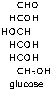
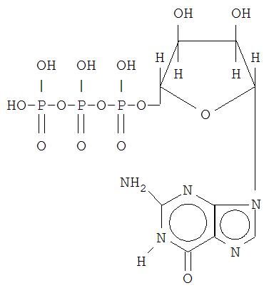
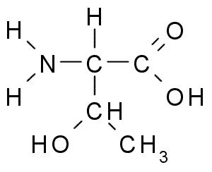
Glucose is an aldohexose - the natural form, D-glucose is also called dextrose. Glucose is biologically important, and is one of the products of photosynthesis by plants and some prokaryotes. Glucose is an important precursor in the synthesis of glycogen (animals), polysaccharides, lactose, cellulose and starch (plants), proteins and lipids, and vitamin C (plants, most animals).

Glucose is an important fuel in energy yielding catabolic pathways such as glycolysis, the pentose phosphate pathway, and the citric acid cycle. In the catabolic pathway gluconeogenesis, animals convert non-carbohydrate intermediates such as pyruvate and glycerol into glucose. Animals and fungi convert glycogen to glucose (glycogenolysis), while plants hydrolyse the storage form starch into glucose.

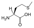
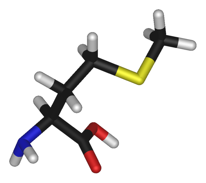
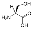
Glycosylation involves the addition of saccharides to proteins and lipids, and is one of four principal co-translational and post-translational modification steps in the synthesis of membranous and secretory proteins. Most proteins synthesized in the rough ER undergo glycosylation, which is an enzyme-directed site-specific process, as opposed to the non-enzymatic chemical reaction of glycation (below). There are two types of glycosylation: N-linked glycosylation to the amide nitrogen of asparagine side chains and O-linked glycosylation to the hydroxy oxygen of serine and threonine side chains.

Glucose can form formaldehyde under abiotic conditions, a propery that probably was utilized by primitive biochemical systems. A more important property of glucose to advanced metabolic forms is the low tendency of glucose, in comparison to other hexose sugars, to react non-specifically with the amino groups of proteins. This glycation reaction reduces or destroys the function of many enzymes. Glucose's low rate of glycation is due to the tendency of glucose to adopt the less reactive cyclic isomeric form (below). Nevertheless, many of the long-term complications of diabetes, such as blindness, kidney failure, and peripheral neuropathy probably result from the glycation of proteins or lipids.

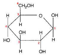
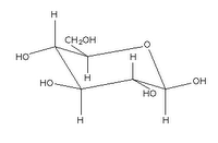
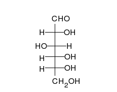
Glucose can take up a linear or a cyclic form. The ring form results from bonding between the aldehyde C atom and the C-5 hydroxyl group, creating an intramolecular hemiacetal. By virtue of the ring form's resemblance to pyran, glucose is also termed glucopyranose.

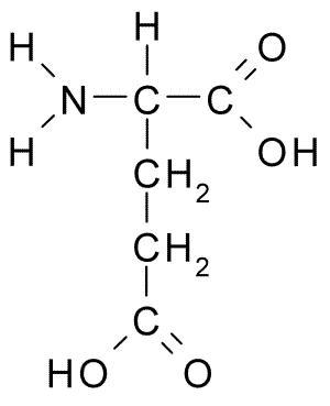
Glucose has 4 optic centers, so glucose has (4²-1) = 15 possible optical stereoisomers, of which only 7 occur biologically. Of these galactose (Gal) and mannose (Man) are the most important. The eight isomers (including glucose) are all diastereoisomers in relation to each other and all belong to the D-series.

An additional asymmetric center at C-1 (called the anomeric carbon atom) is created when glucose cyclizes, so two ring structures, called anomers, α-glucose and β-glucose. The α-and β- forms differ structurally in the orientation of the hydroxyl group linked to C-1 in the ring. The designation α means that the C-1 hydroxyl group is below the plane of the ring in the Haworth projuection, while the β means it is above the ring. The α and β forms slowly interconvert in aqueous solution in a process called mutarotation solution to a final stable ratio of α:β = 36:64.

Image of glucose representations from Fisher to Haworth.
Animated image of D-Glucose shifting into β-D-glucose.
. . . since 10/06/06